A TIPIC Ultrasonographic B-Mode Imaging of the Common Carotid
4.5 (735) · $ 24.50 · In stock
Introduction/Patient Description Extracranial carotid duplex ultrasonography (DUS) was requested within 2 weeks after sudden onset of unilateral, evolving, neck pain. Signs and symptoms related to a 53 year-old man included local swelling, skin changes, increased, local sensations, and high sensitivity to palpation. Atherosclerotic risk factors were not noted. He had contralateral radiation therapy, neck and

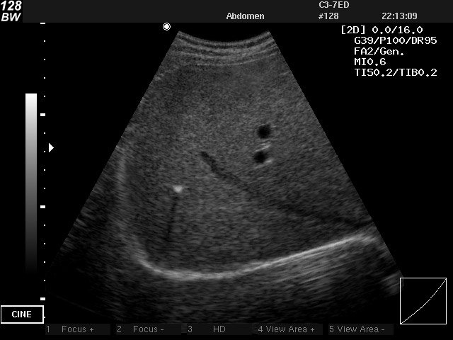
B-mode ultrasound image of a longitudinal section of the carotid artery
Frontiers High-Frame Rate Vector Flow Imaging Technique: Initial Application in Evaluating the Hemodynamic Changes of Carotid Stenosis Caused by Atherosclerosis
Physics of Ultrasound - NYSORA
Neuro-ultrasound - EMCrit Project

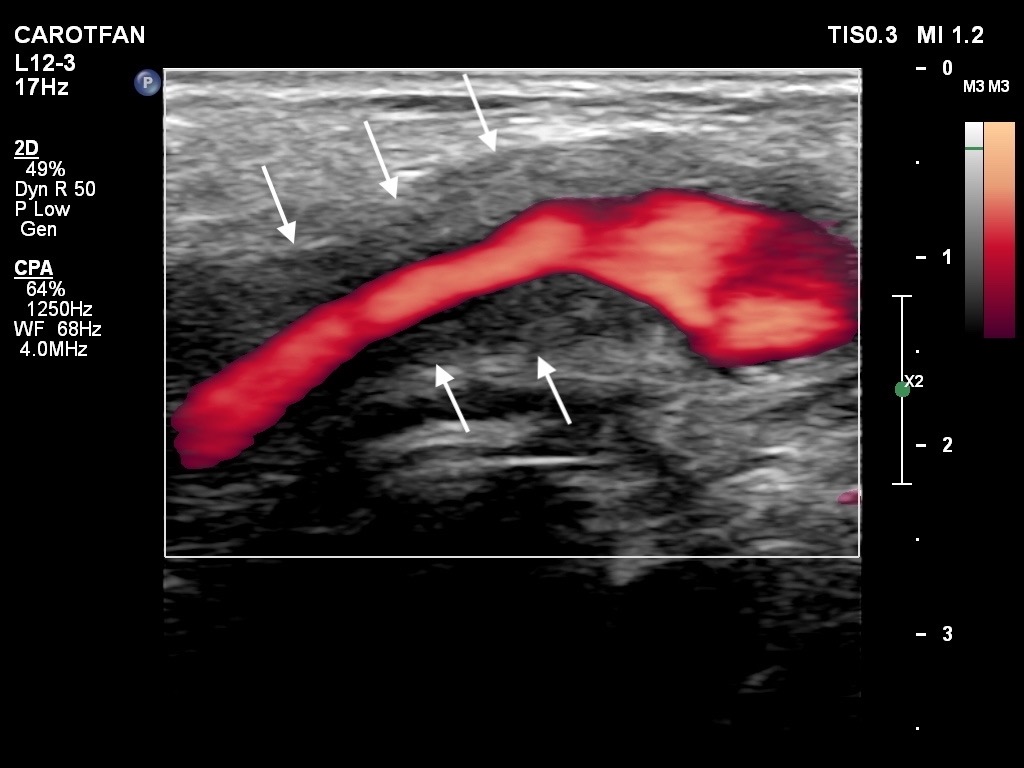
B-mode ultrasound image of the common carotid artery (CCA): a border of

SVU 45th Annual Conference: Vascular Imaging Educators Workshop

Development of a Duplex Ultrasound Simulator and Preliminary Validation of Velocity Measurements in Carotid Artery Models - R. Eugene Zierler, Daniel F. Leotta, Kurt Sansom, Alberto Aliseda, Mark D. Anderson, Florence H.

Colour Doppler evaluation of extracranial carotid artery in patients presenting with features of cerebrovascular disease: A clinical and radiological correlation
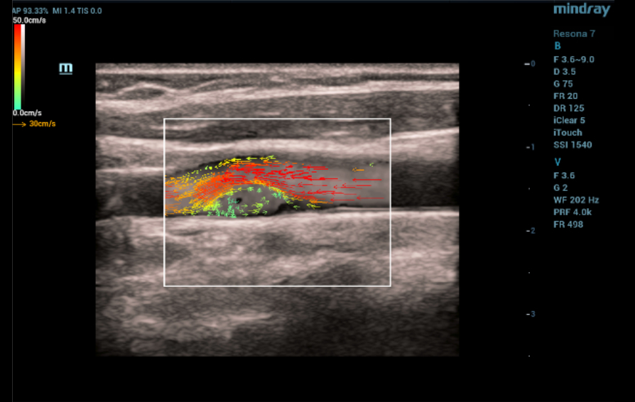
Ultrasound Journal 13 - Ultrasound Diagnostics with Carotid-Web Using V Flow Technology - Mindray
Doppler ultrasound of carotid arteries

Cerebrovascular Sonography - Lange Review Ultrasonography Examination, 4th Edition

PDF) B-mode ultrasound common carotid artery intima-media thickness and external diameter: cross-sectional and longitudinal associations with carotid atherosclerosis in a large population sample