Optical Coherence Tomography: Imaging Mouse Retinal Ganglion Cells In Vivo
4.9 (224) · $ 14.50 · In stock

Scientific Article | Structural changes in the retina are common manifestations of ophthalmic diseases.
Fundus photography, fluorescein angiography, optical coherence tomography and electroretinography of preclinical animal models of ocular diseases - Kumar - Annals of Eye Science

PDF) Srgap2 suppression ameliorates retinal ganglion cell degeneration in mice
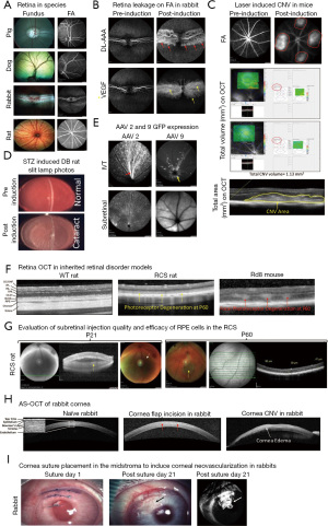
Frontiers Characterization of the Canine Retinal Vasculature With Optical Coherence Tomography Angiography: Comparisons With Histology and Fluorescein Angiography

Transplanted human induced pluripotent stem cells- derived retinal ganglion cells embed within mouse retinas and are electrophysiologically functional - ScienceDirect

Correction-free remotely scanned two-photon in vivo mouse retinal
Applied Sciences, Free Full-Text
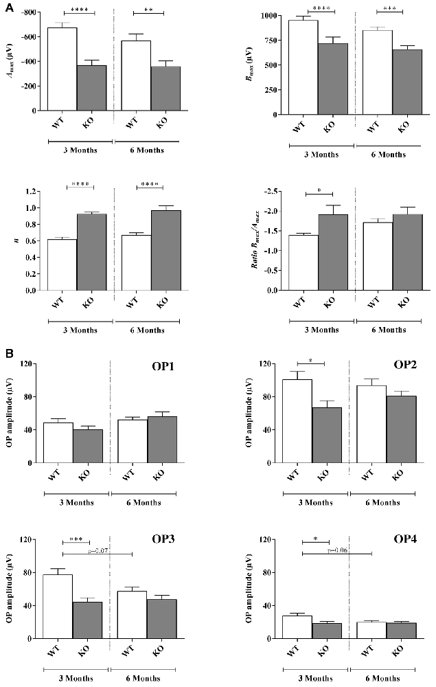
Frontiers Early Retinal Defects in Fmr1−/y Mice: Toward a Critical Role of Visual Dys-Sensitivity in the Fragile X Syndrome Phenotype?

Image segmentation of mouse eye in vivo with optical coherence

Retina imaging — Klinisches Sensoring und Monitoring — TU Dresden
Frontiers Topical nerve growth factor prevents neurodegenerative and vascular stages of diabetic retinopathy
PDF) Early Retinal Defects in Fmr1−/y Mice: Toward a Critical Role of Visual Dys-Sensitivity in the Fragile X Syndrome Phenotype?

C57Bl/6 retinal scans and posterior segmental layer thi