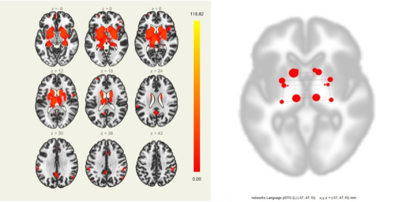
Coronal and axial slices displaying the IFG area that showed
4.7 (735) · $ 25.00 · In stock
EPOS™

David ZALD, Professor (Full), Ph.D.

Dissociating the white matter tracts connecting the temporo-parietal cortical region with frontal cortex using diffusion tractography. - Abstract - Europe PMC

STN Stimulation Alters Pallidal—Frontal Coupling during Response Selection under Competition - Stéphane Thobois, Gary R Hotton, Serge Pinto, Leonora Wilkinson, Patricia Limousin-Dowsey, David J Brooks, Marjan Jahanshahi, 2007
The Neurological Basis of Developmental Dyslexia and Related Disorders: A Reappraisal of the Temporal Hypothesis, Twenty Years on – MéloDys
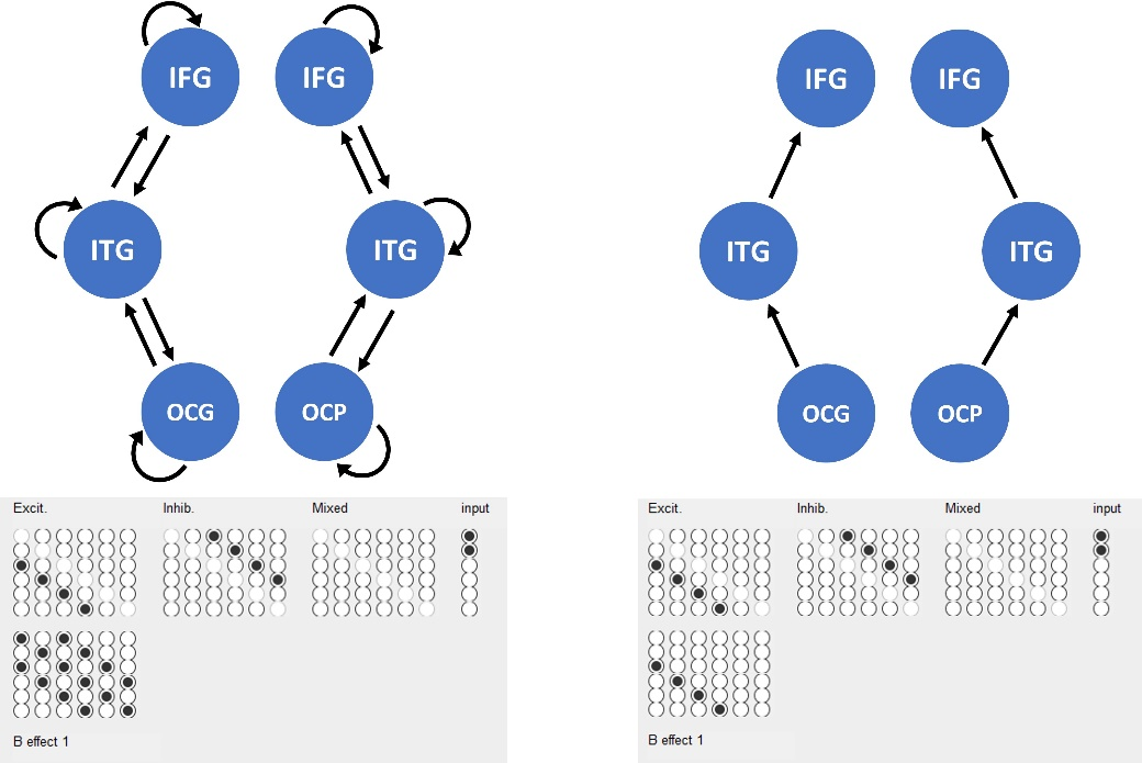
DCM for Evoked Responses - SPM Documentation
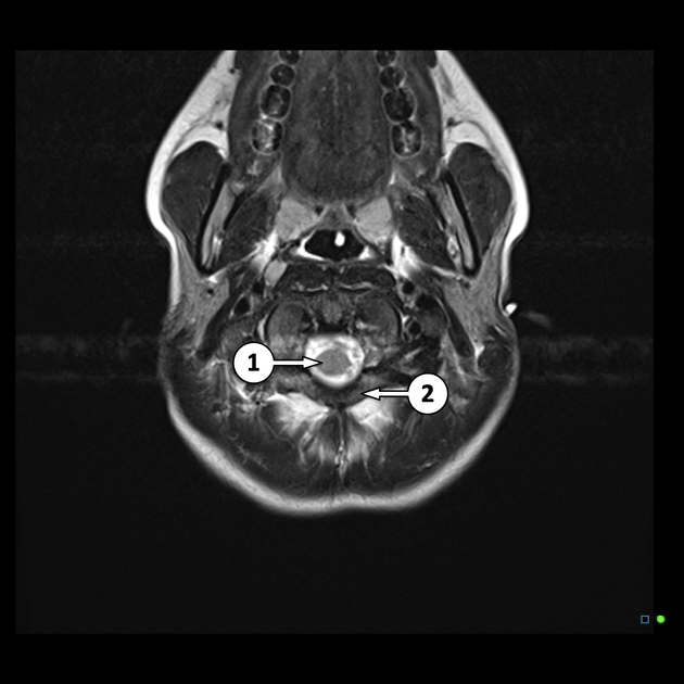
MRI head axial T2 - labeling questions, Radiology Case

An atlas of white matter anatomy, its variability, and reproducibility based on Constrained Spherical Deconvolution of diffusion MRI

Matthew C Hagen's research works University of Minnesota Duluth

Axial view of left dorsal AF across all subjects in gre
State of Brain Networks in Long Covid: Post Mild to Moderate Acute Covid -19 Disease
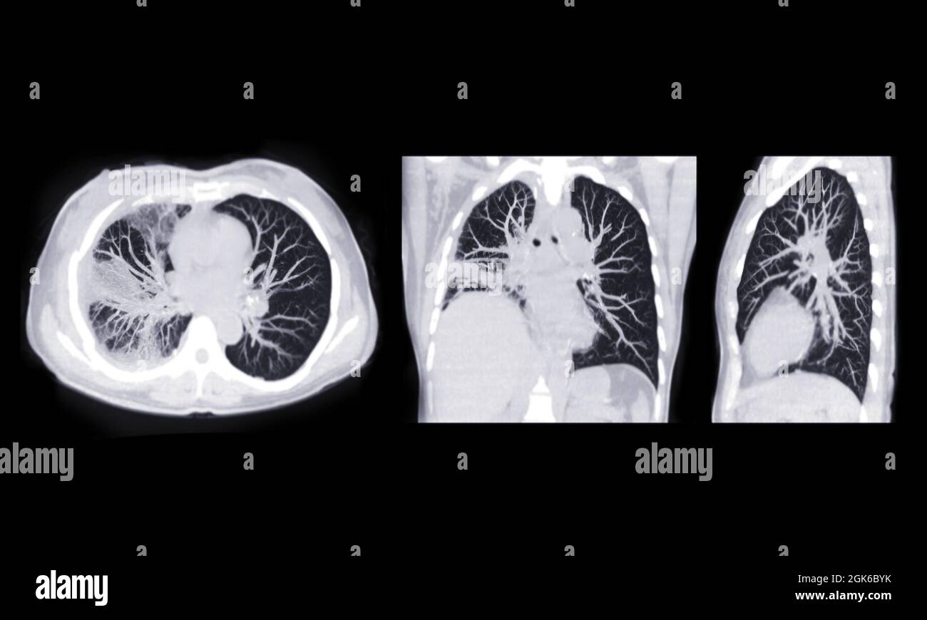
CT scan of Chest or lung axial, coronal and sagittal mip view of lung infection covid-19 with ground glass opacity Stock Photo - Alamy
Coronal, sagittal, and axial slices showing the amygdala seed region
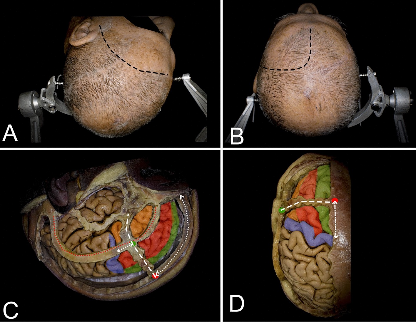
Cortical and white matter anatomy relevant for the lateral and superior approaches to resect intraaxial lesions within the frontal lobe