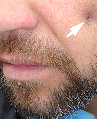
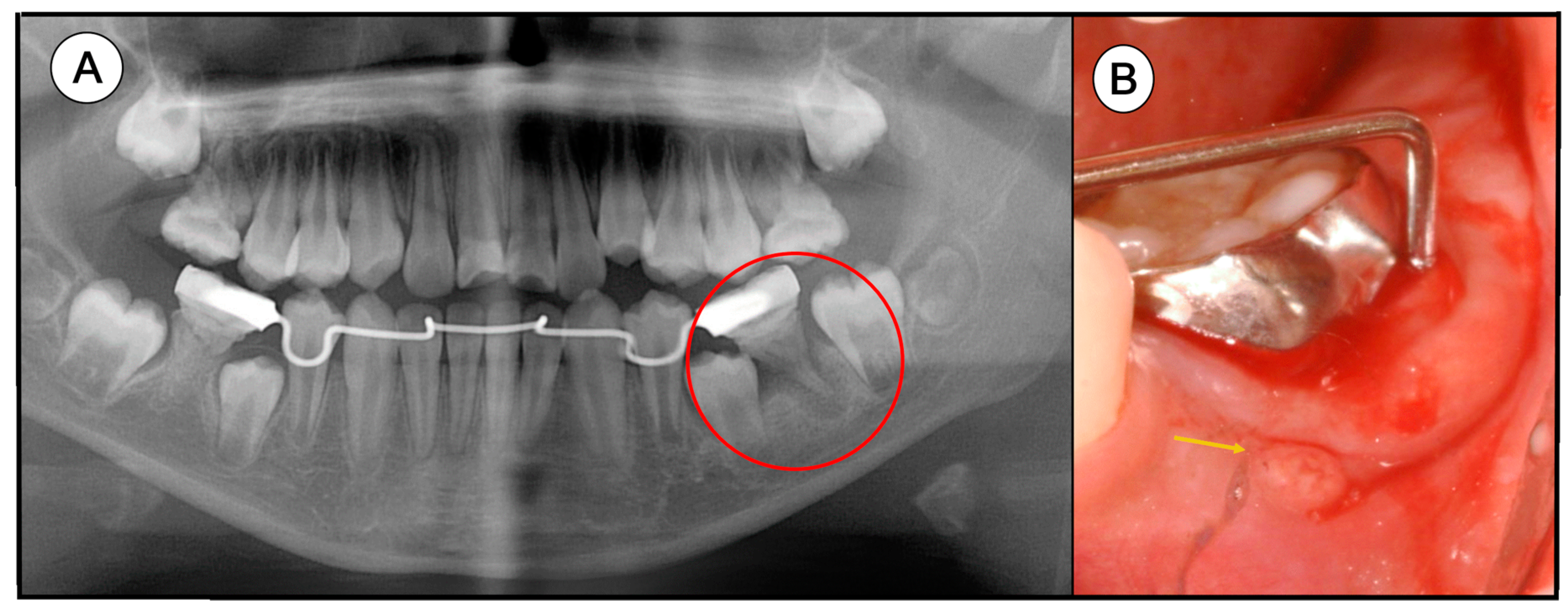
a Mandibular fistula indicated by an arrow in the apical region of dd
4.6 (724) · $ 31.00 · In stock
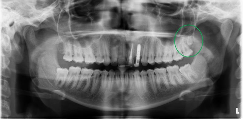
Download scientific diagram | a Mandibular fistula indicated by an arrow in the apical region of dd 36-37. b A fistula in the apical region of dd 46-47 (white arrows) and a red area in the mucosa (black arrows) are seen in the right lingual surface of the mandible. c Panoramic radiograph showing no bone lesions in the mandible. d Periapical x-ray with no bone involvement in the apical region of dd 46-47 from publication: Treatment of bisphosphonate-induced osteonecrosis of the jaws with Nd:YAG laser biostimulation | Osteonecrosis, Jaw and Nd:YAG Laser | ResearchGate, the professional network for scientists.

Dental CT: Pathologic Findings in the Teeth and Jaws
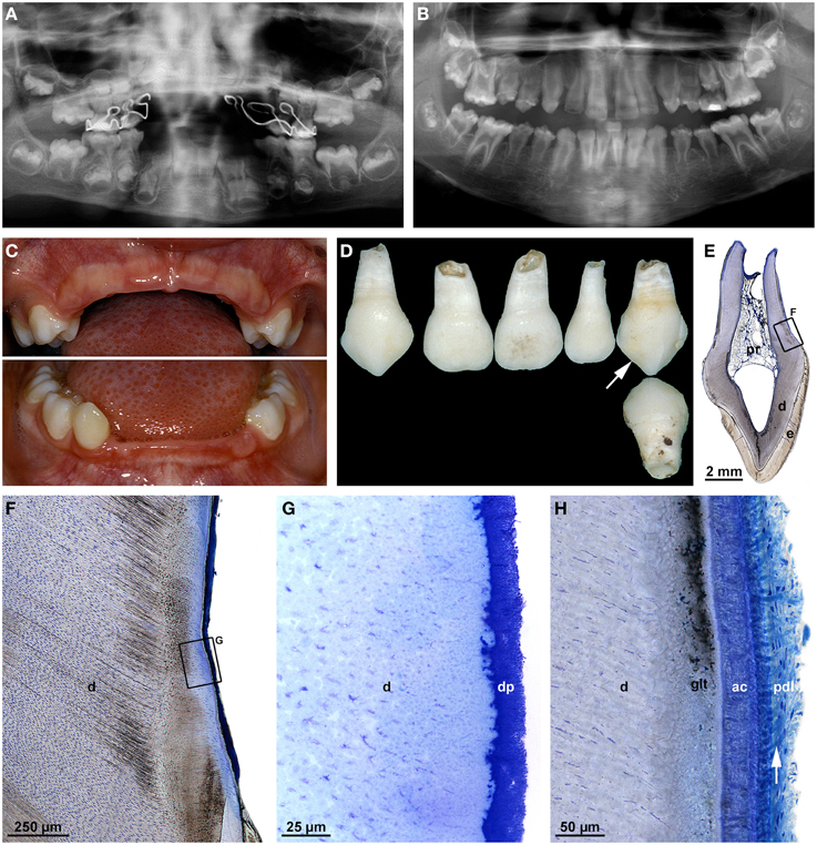
Frontiers Malformations of the tooth root in humans

Satu ALALUUSUA, University of Helsinki, Helsinki, HY, Institute of Dentistry

Radiolucent lesions of the mandible: a pattern-based approach to diagnosis, Insights into Imaging
Ilan Rotstein, John I. Ingle, 2019, Ingle's Endodontics Publisher: PMPH USA by Sociedad de Medicina Oral Internacional - Issuu

PDF) Regional Odontodysplasia Affecting the Maxilla

Healthcare, Free Full-Text

SciELO - Brazil - Differential diagnosis and clinical management
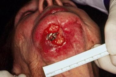
Oral Cutaneous Fistulas: Practice Essentials, Pathophysiology
Case Archive, School of Dental Medicine
![Comparison between hydroxyapatite and polycaprolactone in inducing osteogenic differentiation and augmenting maxillary bone regeneration in rats [PeerJ]](https://dfzljdn9uc3pi.cloudfront.net/2022/13356/1/fig-4-full.png)
Comparison between hydroxyapatite and polycaprolactone in inducing osteogenic differentiation and augmenting maxillary bone regeneration in rats [PeerJ]
Single and Multiple Odontogenic Cutaneous Sinus Tracts
Applied Sciences, Free Full-Text

a Mandibular fistula indicated by an arrow in the apical region of dd
:max_bytes(150000):strip_icc()/rs-best-places-to-buy-swimsuits-tout-c5ed2ec2b1de428ba4cbefaaedd06a9b.jpg)