A) Preoperative intraoral periapical (IOPA) radiograph of 36. B
4.5 (466) · $ 26.50 · In stock

A) Preoperative intraoral periapical (IOPA) radiograph of 36. B) Post operative (IOPA) radiograph of 36. C) 1 month follow up IOPA radiograph of 36. D) 6 months follow up IOPA radiograph of 36. E) 1 year follow up IOPA radiograph of 36. - IP Indian J Conserv Endod - clinical and preclinical conservative /restorative de

A short-term prospective study to evaluate the

a) Preoperative IOPA radiograph of tooth #36. (b) Intraoral image

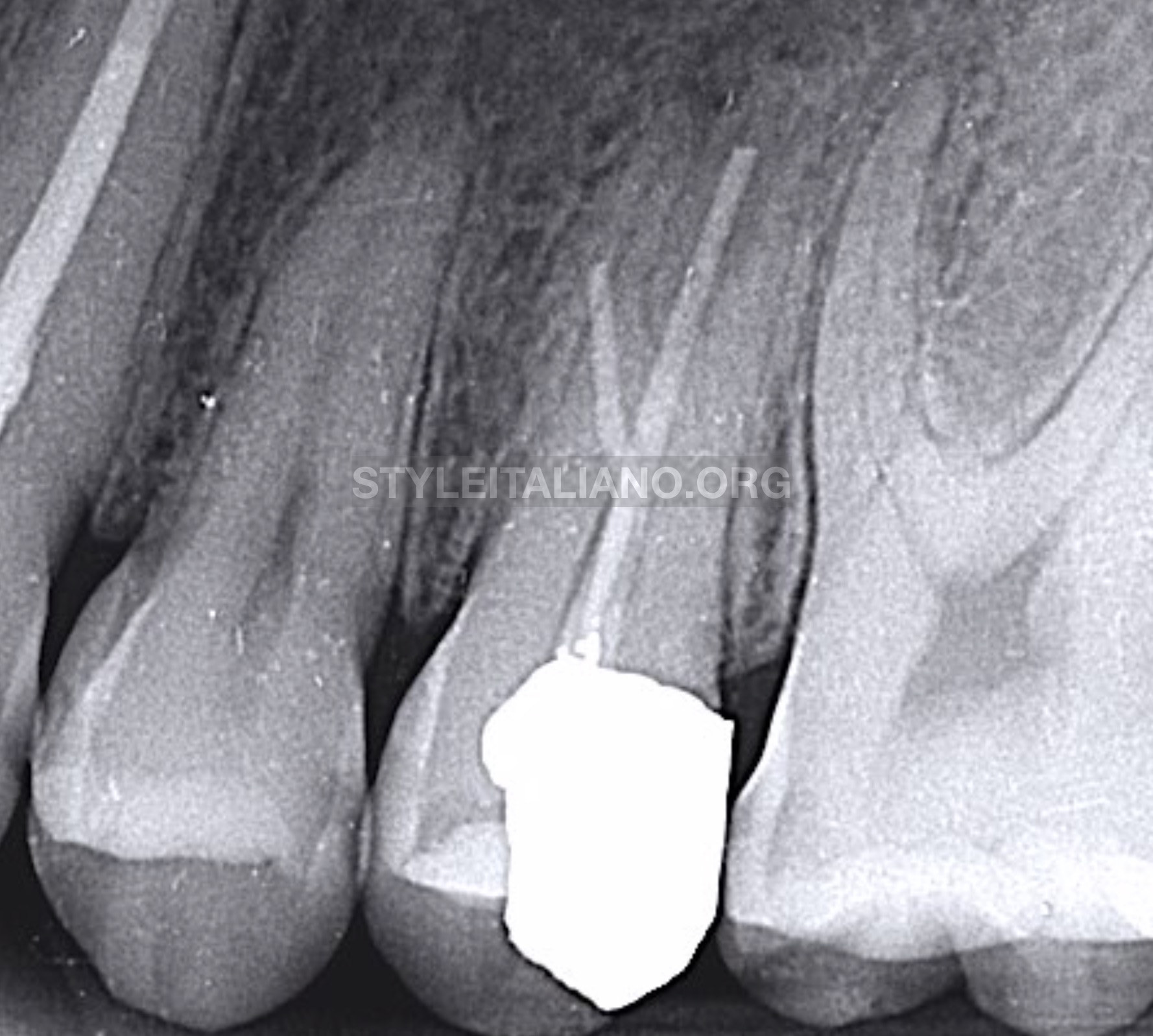
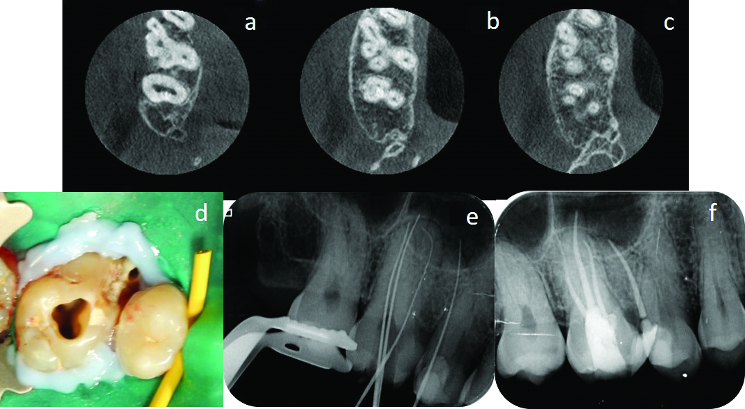
Endodontic management of a maxillary first molar with three roots

Radiology of Apical Periodontitis - Essential endodontology
The advantages of pre-operative radiograph in the diagnosis and in

a) IOPA radiograph after obturation, buccal view. (b) IOPA
jcdr-13-ZD01-g002.jpg

Radiographic findings. (a) Straight on angulation; (b) Mesial

A) Preoperative intraoral periapical (IOPA) radiograph of 36. B

PDF) Direct pulp capping with bioactive materials – A case series

Postoperative IOPA of 36 and 46.
![PDF] Image Processing on IOPA Radiographs: A comprehensive case](https://d3i71xaburhd42.cloudfront.net/a7e90979a1332cfc2e8c8cda0757b00ec195ecf2/7-Figure17-1.png)
PDF] Image Processing on IOPA Radiographs: A comprehensive case

Nonsurgical Management of Periapical Lesion: A Case Series