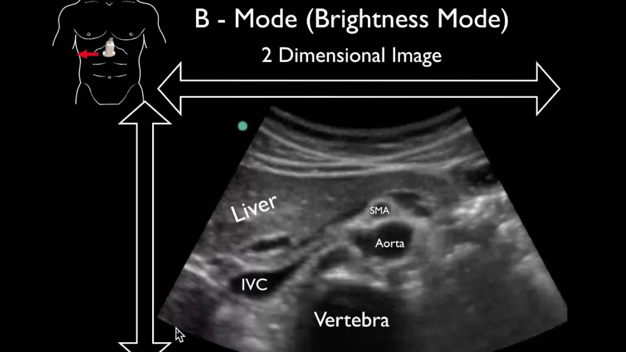
A) A brightness mode (b-mode) image of the lateral abdominal wall.
4.8 (601) · $ 20.99 · In stock

Download scientific diagram | (A) A brightness mode (b-mode) image of the lateral abdominal wall. Abbreviations: EO, external oblique; IO, internal oblique; TrA, transversus abdominis. (B) A split-screen image with b-mode on the left and motion mode (m-mode) on the right. The m-mode image represents the information from the dotted line on the b-mode image displayed over time (x-axis). Static structures produce straight interfaces while structures that change in thickness or depth (in this case the TrA) create curved interfaces. The increase in depth of the TrA correlates to a contraction. Reproduced with permission Whittaker 2007. 142 from publication: Rehabilitative Ultrasound Imaging: Understanding the Technology and Its Applications | The use of ultrasound imaging by physical therapists is growing in popularity. This commentary has 2 aims. The first is to introduce the concept of rehabilitative ultrasound imaging (RUSI), provide a definition of the scope of this emerging tool in regard to the physical | Rehabilitation, Ultrasonography and Ultrasound Imaging | ResearchGate, the professional network for scientists.

Diagnostic point-of-care ultrasound (POCUS) for gastrointestinal pathology: state of the art from basics to advanced, World Journal of Emergency Surgery
Modes Ultrasound A-mode- amplitude mode. B-mode- brightness mode. - ppt video online download

Is tissue harmonic ultrasound imaging (THI) of the prostatic urethra and rectum superior to brightness (B) mode imaging? An observer study - ScienceDirect

Bridging repair of the abdominal wall in a rat experimental model. Comparison between uncoated and polyethylene oxide-coated equine pericardium meshes

PDF) Rehabilitative Ultrasound Imaging: Understanding the

Diagnostics, Free Full-Text

Measurement of linea alba distortion and linea alba stiffness. (A)

Ultrasound Machine Basics-Knobology, Probes, and Modes - POCUS 101

Jackie WHITTAKER, Professor (Associate), BScPT, PhD
![PDF] Ultrasound imaging of the abdominal muscles and bladder: implications for the clinical assessment of individuals with lumbopelvic pain](https://d3i71xaburhd42.cloudfront.net/9cdb06554c0b6a8a478a96007716c5f0268a194b/347-Figure9-1.png)
PDF] Ultrasound imaging of the abdominal muscles and bladder: implications for the clinical assessment of individuals with lumbopelvic pain