The STL images of two geometries of the 3D-printed bioceramic model
4.8 (227) · $ 10.50 · In stock

Download scientific diagram | The STL images of two geometries of the 3D-printed bioceramic model were designed as follows: The cylindrical compression sample (a), the concave-topped disk structures views of the bottom (c), and the top (d). The cross-section views of concave-top disk structures also showed the STL image of a horizontal section (e) and a vertical section (f). Furthermore, the two kinds of 3D-printed sintered bioceramic images were obtained. The 3D cylinder bioceramic sample (b), the bottom view (g), and the top view (h) of the concave-top disc structure of the 3D-printed bioceramic scaffold from publication: Bilayer osteochondral graft in rabbit xenogeneic transplantation model comprising sintered 3D-printed bioceramic and human adipose-derived stem cells laden biohydrogel | Reconstruction of severe osteochondral defects in articular cartilage and subchondral trabecular bone remains a challenging problem. The well-integrated bilayer osteochondral graft design expects to be guided the chondrogenic and osteogenic differentiation for stem cells and | Bioceramics, Osteochondritis and Grafts | ResearchGate, the professional network for scientists.

Preparation procedures and primary characterization of porous
SiOC ceramics with ordered porosity by 3D-printing of a preceramic
.JPG)
Leveraging Additive Manufacturing to Improve Joint Replacement

5792 PDFs Review articles in NANO-SILICA

Three-dimensional printing of biomaterials for bone tissue

An osteochondral defect was generated in the rabbit knee
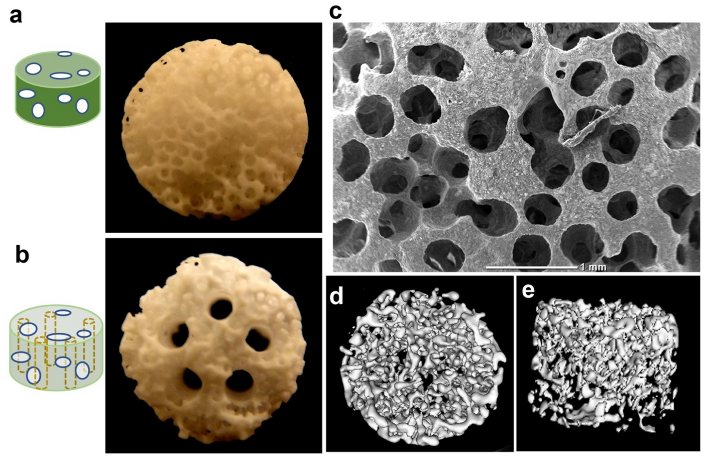
Multiple channels with interconnected pores in a bioceramic

Surface modification of stereolithography-based 3D printed

PDF) Vascularized Bone Tissue Engineering: Approaches for

5792 PDFs Review articles in NANO-SILICA
pub.mdpi-res.com/jfb/jfb-09-00017/article_deploy/h