A) MRI findings: the typical bunched medial collateral ligament (MCL)
4.7 (490) · $ 15.00 · In stock

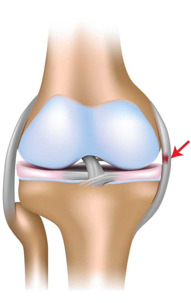
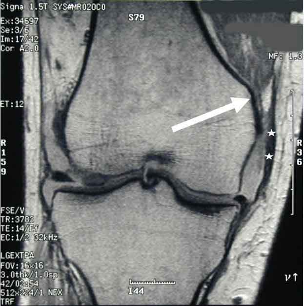
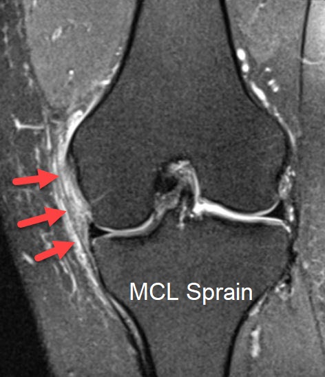
Download scientific diagram | (A) MRI findings: the typical bunched medial collateral ligament (MCL) fibres are obvious on the T2-weighted MR image (arrow). Countercoup oedema is evident in the lateral tibial plateau. (B) Anatomical findings: the fibres are short and abruptly jump over the semitendinosus tendon. The femoral insertion site remained intact. Note. sMCL, superficial MCL. from publication: Isolated medial collateral ligament tears: An update on management | Tears of the medial collateral ligament (MCL) are the most common knee ligament injury. Incomplete tears (grade I, II) and isolated tears (grade III) of the MCL without valgus instability can be treated without surgery, with early functional rehabilitation. Failure of | Tears, Collateral Ligaments and Reconstruction | ResearchGate, the professional network for scientists.
MCL Injury, Medial Collateral Ligament Tear
Medial Collateral Ligament of the Knee - Physiopedia

C.A. ENCINAS-ULLÁN, Medical Doctor, Hospital Universitario La Paz, Madrid, Servicio de Cirugía Ortopédica y Traumatología

Medial Collateral Ligament Injury Of The Knee Mcl Coronal Pd Fat

Medial collateral ligament injury of the knee: correlations

Axial MRI at height of the physis and corresponding line drawing of the

Collateral Ligament Injury MRI: Practice Essentials, Radiography

A) MRI findings: the typical bunched medial collateral ligament (MCL)

PDF) Isolated medial collateral ligament tears: An update on management
MCL Tear, Indiana Slip & Fall Lawyers
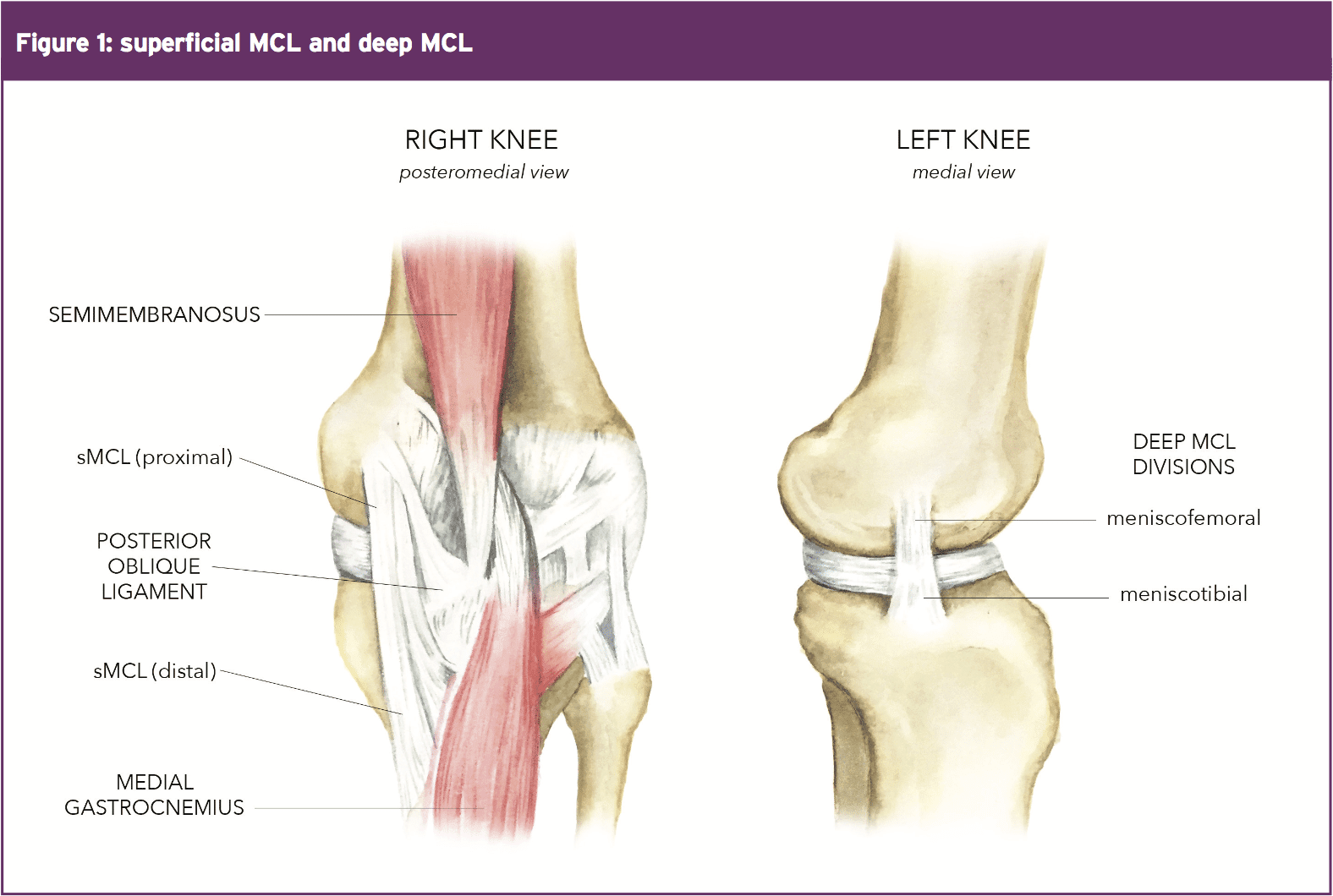
MCL: Anatomy, Biomechanics & Injury Science

PDF) Isolated medial collateral ligament tears: An update on management
How to diagnose and treat a medial collateral ligament and lateral